Dental Erosion

Key Points

  • Dental erosion is a chemical process characterized by acid dissolution of dental hard tissue not involving acids of bacterial origin.
  • Erosive demineralization can result in progressive, irreversible loss of tooth mineral substance, and may be caused by intrinsic (e.g., acid reflux and excessive vomiting) and/or extrinsic (e.g., dietary) factors.
  • Frequent consumption of soft drinks, particularly carbonated sodas, is a primary risk factor for erosive tooth wear. Consumption of acidic snacks/sweets or natural acidic fruit juice may also increase risk for erosion.
  • Diagnosis and management of dental erosion includes careful clinical examination and evaluation of the patient to identify common signs of erosion (e.g., loss of enamel texture, cupping or flattening on occlusal surfaces), predisposing factors for erosive tooth wear, and options to reduce probability of exposure to erosive (acidic) drinks, dietary sources and/or other acids of intrinsic or extrinsic origin.
Introduction

Dental erosion, also known as tooth erosion, is the chemical loss of mineralized tooth substance caused by exposure to acids not derived from oral bacteria.1 Erosive demineralization is a chemical process characterized by acid dissolution of dental hard tissue, and its etiology is multifactorial.2-6

The primary etiologic factors of dental erosion are acids of intrinsic (often due to acid reflux) or extrinsic origin (diet, particularly carbonated/soft drinks or acidic fruit-juice consumption; exposure to industrial or environmental chemicals).3, 4, 7, 8 Beyond aesthetic consequences and associated oral health issues, severe erosive activity can lead to exposed dentin, hypersensitivity, and eventual loss of affected teeth.9-12

Dental erosion can occur in individuals of any age.13, 14 One global prevalence study estimated that the mean prevalence of erosion in deciduous teeth ranges between 30% and 50%, and the mean prevalence of erosion in permanent teeth in adults ranges between 20% and 45%.14 The same study also reported selective and highly variable data on erosion prevalence in children, adolescents and adults, due to variation in erosion indices, populations examined, regional diets/customs and study design.14 A 2015 meta-analysis13 indicated that 34.1% of 16,661 children and adolescents worldwide exhibited dental erosion. In the U.S., an analysis of data from the National Health and Nutrition Examination Survey (NHANES, 2003-2004) estimated an erosive tooth wear prevalence of 45.9% among children15 and up to 80% among adults.16

Dental Erosion

Intrinsic Causes

Intrinsic erosion results from the introduction of gastric acids into the oral cavity at a frequency, duration and/or intensity that exceed the ability of the buffering capacity of saliva or other oral health measures to minimize an erosive challenge, usually several times a week for an extended period of time.7, 13  Stomach acid may reach the oral cavity in cases of gastroesophageal reflux disease (GERD), a common condition in which gastric contents reflux back up into the esophagus and/or the mouth. Other erosive challenges from gastric acid occur from recurrent vomiting, such as occurs in bulimia nervosa, chronic alcoholism, and pregnancy, when it is referred to as hyperemesis gravidarum.2, 4, 7, 17

  • Gastroesophageal reflux. Gastroesophageal reflux disease is considered a predisposing factor for dental erosion due to chronic regurgitation of gastric contents. Some systematic reviews report that severity of erosive tooth wear may be associated with the frequency and/or intensity of acid regurgitation.18-20 Occasional regurgitation of stomach acids following meals, especially after overeating, is considered normal7, 13 for up to about one hour a day.4 However, in people with GERD, the passage of gastric acids into the oral cavity during sleep is especially damaging to the teeth, as salivation and swallowing are reduced, and, in a supine position, the lower molars can be bathed in the acids.4 Increases in intra-abdominal pressure (i.e., from obesity or pregnancy) may also increase reflux,7 and GERD has been found to be a common comorbidity of several chronic respiratory conditions.21-23 

    Two common symptoms of GERD are heartburn and/or acid regurgitation, and additional manifestations of GERD include dysphagia, dyspepsia, hoarseness of voice, abdominal pain, or an acidic taste (or burning sensation) in the mouth or throat.24-27 Dysregulation of gastrointestinal enzymes and hormones and impaired reflux clearance have been identified as two potentially important contributors to GERD development.28, 29

    Prevalence rates of GERD across all age groups reportedly ranges from 9% to 33%, with some evidence suggesting an increase in GERD prevalence in the United States since 1995.30 One systematic review determined that the mean prevalence of erosion in adult patients with GERD was 32.5% (range: 21-83%) and 17% among children with GERD (range: 14-87%).31 Additionally, one meta-analysis concluded that individuals with at least weekly symptoms of GERD have up to five times higher risk of esophageal adenocarcinoma.32   
     

Figures, below. Moderate to severe erosion from GERD in the primary dentition.

erosion molarserosion molars
Photos courtesy of Martha Ann Keels, D.D.S., PhD
 
  • Bulimia. Bulimia nervosa is a serious eating disorder that is characterized by self-induced vomiting as a means to maintain a desired weight.4, 7, 33 It is a relatively common disorder among women in Western industrialized nations, with a prevalence of approximately 5% in 18-35 year-old females.7 Among individuals with bulimia, the prevalence of erosion has been reported to be over 90%.7 Individuals with bulimia or other eating disorders are commonly found to have poor oral health, with one systematic review finding that patients with eating disorders and/or self-induced vomiting had five to seven times higher risk of dental erosion.34 Because patients with bulimia are generally of average weight, dentists are often the first to recognize the condition by the characteristic erosion of the lingual-palatal aspect of the anterior maxillary teeth (i.e., perimolysis or perimylolysis, see photos below),4, 7 caused by the forceful expulsion of stomach acids onto the front teeth during vomiting.

Figures, below. Characteristic erosion pattern from bulimia.

Exterior view of enamel erosion of the lingual-palatal aspect of the anterior maxillary teeth (i.e., perimolysis or perimylolysis)Internal view of enamel erosion of the lingual-palatal aspect of the anterior maxillary teeth (i.e., perimolysis or perimylolysis)
Photos courtesy of Craig Mabrito, D.D.S.
 
  • Chronic Alcoholism. Alcoholism, with a prevalence of around 10% in Western countries,7 can result in erosive tooth wear when there is frequent vomiting and/or increased regurgitation7, 35 or even when alcohol consumption is low-dose but long-term.36 In addition, erosion in individuals with alcoholism can also be exacerbated by the consumption of acidic drinks.10, 17 Small studies have found between 49 – 92% of the teeth in individuals with alcoholism show signs of erosion.7, 36 As in bulimia, the palatal surfaces of anterior maxillary teeth tend to be the most affected in alcoholic patients, who were also found to have relatively high erosion severity on the occlusal and palatal surfaces of the posterior, and incisal edges of the anterior, maxillary teeth.7, 35, 36

  • Pregnancy. Although dental erosion is rare in pregnancy, it has been reported in women who experience hyperemesis gravidarum,7 where the nausea and vomiting may be experienced over a longer period of time and may be more severe.37

Extrinsic Causes

Erosion due to extrinsic factors may arise from a combination of dietary, lifestyle, environmental or occupational factors that expose teeth to acids in beverages2, 3, 5 or inorganic acid vapors in the environment.2, 5

  • Beverages. A growing body of evidence suggests that the primary predisposing factor for extrinsic dental erosion2, 4, 13 is frequent consumption of soft drinks, sports drinks and fruit juices with low pH values (2.0-3.5).4, 5 Any beverage with a low pH can increase risk for erosion,4 especially with frequent consumption.2, 4, 5, 17, 38Although wines, for example, are naturally acidic, they are not commonly cited as a risk factor for erosion because they are typically not consumed as frequently as sports drinks and diet sodas.5 In 2016, a study by Reddy et al. measured the pH of 379 commercially available beverages available in the U.S., and found that 93% had a pH of less than 4.0.39

    A number of studies6, 13, 17, 40 support the hypothesis that dietary acids are a major contributing factor in dental erosion.7, 41 A 2012 meta-analysis found more than double the risk of erosion from soft drinks as well as an increase in risk from chewable vitamin C tablets.40 Other studies, including a 2015 meta-analysis, determined that natural fruit juices as well as carbonated soft drinks and acidic, sweet snacks and sweet-sour candies were significant predisposing factors for erosion.13, 42

  • Lifestyle. More frequent consumption of highly acidic fruit and sport drinks, in combination with decreased salivary flow and dehydration from athletic or strenuous activity, may increase erosion risk.3 Intense workouts may also increase the possibility of gastroesophageal reflux.3 Although there is some evidence that a vegetarian diet and excessive use of vinegar-based dressings may lead to increased erosion,43-45 a critical analysis of this topic noted that the evidence on erosivity of vegetarian diets has significant limitations and is relatively weak in overall quality.46

  • Environmental and Occupational Risks. Airborne industrial acids have been implicated in dental erosion among factory workers,2, 3 particularly in munitions, battery, and fertilizer plants.3 Swimmers frequenting chlorinated swimming pools have been reported to have increased levels of erosion, attributed to the water’s pH.2, 3, 10

Diagnosis, Prevention, and Management

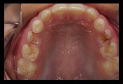
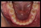
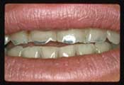
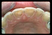
Erosive tooth wear may be diagnosed by integrating a review of findings from a detailed health history, assessment of relevant risk factors, and a comprehensive clinical examination.1 The process of erosive tooth wear begins with initial softening of the enamel surface with subsequent and/or progressive loss of volume, with a softened layer (i.e., less surface hardness) remaining at the mineralized tooth surface.47 Individuals with erosion may also present with dentition that has loss of enamel texture and/or a silky, glossy or “melted” appearance, with cupped, flattened or smoothed occlusal surfaces (or without the sound morphology of natural dentition).1, 27,48

A detailed health history-taking process can help identify high-risk conditions (e.g., GERD, bulimia) or underlying causes that can elevate risk of exposure to acid erosive challenges, primarily acids of intrinsic (gastric) or extrinsic (dietary) origin. In the oral examination, the dentist may observe common signs of erosive tooth wear, such as shallow defects (typically on smooth surfaces) and cupping or flattening on occlusal surfaces (examples presented in photos in the “Gastroesophageal reflux” section above).1 Additional signs of progressive erosive tooth wear may include dentin hypersensitivity and/or erosive lesions with absence of staining.27, 49 Early diagnosis of dental erosion can be challenging because patients often present without clear or clinically evident symptoms.27

Since dental erosion results in progressive and irreversible loss of mineralized tooth substance, the primary focus of erosion intervention is prevention and reduction, followed by management.17 Clinical management typically includes lifestyle and dietary changes, and, if necessary, restorative treatment to halt progression of erosive lesions.17, 50

Some studies have found that milk and yogurt products (presumably unsweetened) may have a protective effect against erosion13, 17, 40 hypothesized to be due to their calcium and phosphate content.40 While calcium supplemented to acidic beverages, and other calcium-enriched products, have been suggested as protective alternatives to soft and sports drinks,17, 40 the efficacy of this strategy has not been thoroughly investigated. Fluoride may contribute to remineralization of enamel,9, 51 but others have advised that fluoride’s “ability to prevent erosion cannot be presumed.”3

More recently, however, several in situ studies52-59 suggest that fluoride treatments are effective in protecting dental enamel from the effects of erosion, although efficacy varies according to compound or preparation, and further research is needed.

Patient Information
  • Avoid dietary acids between meals, reduce intake of acidic beverages and eliminate behaviors that increase acidic (erosive) challenges to the dentition, particularly swishing or straining the liquid between the teeth, or holding the liquid in the mouth.3, 17, 58, 60 Drinking with a straw positioned behind the front teeth can minimize bathing the teeth.17
  • Drink water while eating, or rinse the mouth with water after consuming acidic drinks, candies, or foods.4, 40
  • After vomiting, rinse the mouth with water, a sodium bicarbonate rinse, or milk.4, 40, 61, 62
  • Saliva helps buffer and remove acids; chewing gum may help protect teeth from erosion by promoting salivary flow.
  • Drink milk along with acidic meals or beverages, which contributes to remineralization and helps neutralize acids. 
  • Rinse with water rather than brushing teeth immediately after consuming acidic beverages.4, 13, 63
  • Brush teeth using a soft-bristle brush and fluoride toothpaste.63 Dental abrasion (physical loss of mineralized tooth substance from objects other than teeth) may potentially occur in rare cases of excessive or aggressive toothbrushing. However, normal toothbrushing practices using a soft-bristle brush and low-abrasion, fluoride-containing toothpaste are considered unlikely to cause erosive wear of enamel.49, 64, 65
ADA Seal of Acceptance: Dental Erosion

Look for the ADA Seal of Acceptance—your assurance that the product has been objectively evaluated for safety and efficacy by an independent body of scientific experts, the ADA Council on Scientific Affairs. A company earns the ADA Seal for a product to help prevent or reduce enamel erosion from dietary acids by providing scientific evidence demonstrating the safety and efficacy of its product, which is evaluated according to objective requirements related to the product’s claims.

References
  1. Schlueter N, Amaechi BT, Bartlett D, et al. Terminology of erosive tooth wear: Consensus report of a workshop organized by the ORCA and the Cariology Research Group of the IADR. Caries Research 2020;54(1):2-6.
  2. Imfeld T. Dental erosion. Definition, classification and links. Eur J Oral Sci 1996;104(2 (Pt 2)):151-5.
  3. Zero DT. Etiology of dental erosion--extrinsic factors. Eur J Oral Sci 1996;104(2 (Pt 2)):162-77.
  4. Schlossman M, Montana M. Preventing damage to oral hard and soft tissues. In: Spolarich AE, Panagakos FS, editors. Prevention across the lifespan: A review of evidence-based interventions for common oral conditions. Charlotte: Professional Audience Communications, Inc.; 2017. p. 97-120.
  5. Mandel L. Dental erosion due to wine consumption. JADA 2005;136(1):71-75.
  6. Huysmans MC, Chew HP, Ellwood RP. Clinical studies of dental erosion and erosive wear. Caries Res 2011;45 Suppl 1(Suppl. 1):60-8.
  7. Scheutzel P. Etiology of dental erosion--intrinsic factors. Eur J Oral Sci 1996;104(2 (Pt 2)):178-90.
  8. Saads Carvalho T, Lussi A. Chapter 9: Acidic beverages and foods associated with dental erosion and erosive tooth wear. Monogr Oral Sci 2020;28:91-98.
  9. Twetman S. The evidence base for professional and self-care prevention-caries, erosion and sensitivity. BMC Oral Health 2015;15(1):S4.
  10. Meurman JH, ten Cate JM. Pathogenesis and modifying factors of dental erosion. Eur J Oral Sci 1996;104(2 ( Pt 2)):199-206.
  11. ten Cate J, Imfeld T. Dental erosion, summary. Eur J Oral Sci 1996;104(2):241-44.
  12. Vailati F, Belser UC. Full-mouth adhesive rehabilitation of a severely eroded dentition: The three-step technique. Part 2. Eur J Esthet Dent 2008;3(2):128-46.
  13. Salas MM, Nascimento GG, Vargas-Ferreira F, et al. Diet influenced tooth erosion prevalence in children and adolescents: Results of a meta-analysis and meta-regression. J Dent 2015;43(8):865-75.
  14. Schlueter N, Luka B. Erosive tooth wear - a review on global prevalence and on its prevalence in risk groups. Br Dent J 2018;224(5):364-70.
  15. McGuire J, Szabo A, Jackson S, Bradley TG, Okunseri C. Erosive tooth wear among children in the United States: Relationship to race/ethnicity and obesity. Int J Paediatr Dent 2009;19(2):91-8.
  16. Okunseri C, Wong MC, Yau DT, McGrath C, Szabo A. The relationship between consumption of beverages and tooth wear among adults in the United States. J Public Health Dent 2015;75(4):274-81.
  17. Lussi A, Schlueter N, Schmalz G, et al. Consensus report of the European Federation of Conservative Dentistry. Clinical Oral Investigations 2015;19(7):1557-61.
  18. Ortiz ADC, Fideles SOM, Pomini KT, Buchaim RL. Updates in association of gastroesophageal reflux disease and dental erosion: Systematic review. Expert Review of Gastroenterology & Hepatology 2021:1-10.
  19. Picos A, Badea ME, Dumitrascu DL. Dental erosion in gastro-esophageal reflux disease. A systematic review. Clujul Med 2018;91(4):387-90.
  20. Jordão HWT, Coleman HG, Kunzmann AT, McKenna G. The association between erosive toothwear and gastro-oesophageal reflux-related symptoms and disease: A systematic review and meta-analysis. J Dent 2020;95:103284.
  21. Cazzola M, Segreti A, Calzetta L, Rogliani P. Comorbidities of asthma: Current knowledge and future research needs. Curr Opin Pulm Med 2013;19(1):36-41.
  22. Lee AL, Goldstein RS. Gastroesophageal reflux disease in COPD: Links and risks. Int J Chron Obstruct Pulmon Dis 2015;10:1935-49.
  23. Raghu G, Amatto VC, Behr J, Stowasser S. Comorbidities in idiopathic pulmonary fibrosis patients: A systematic literature review. Eur Respir J 2015;46(4):1113-30.
  24. Vakil N, van Zanten SV, Kahrilas P, Dent J, Jones R. The Montreal definition and classification of gastroesophageal reflux disease: A global evidence-based consensus. Am J Gastroenterol 2006;101(8):1900-20; quiz 43.
  25. Broderick R, Fuchs KH, Breithaupt W, et al. Clinical presentation of gastroesophageal reflux disease: A prospective study on symptom diversity and modification of questionnaire application. Digestive Diseases 2020;38(3):188-95.
  26. Kahrilas P, Yadlapati R, Roman S. Emerging dilemmas in the diagnosis and management of gastroesophageal reflux disease [version 1; peer review: 2 approved]. F1000Research 2017;6(F1000 Faculty Rev):1748.
  27. Donovan T, Nguyen-Ngoc C, Abd Alraheam I, Irusa K. Contemporary diagnosis and management of dental erosion. Journal of Esthetic and Restorative Dentistry 2021;33(1):78-87.
  28. Zachariah RA, Goo T, Lee RH. Mechanism and pathophysiology of gastroesophageal reflux disease. Gastrointestinal Endoscopy Clinics of North America 2020;30(2):209-26.
  29. Gillespie MR, Rai V, Agrawal S, Nandipati KC. The role of microbiota in the pathogenesis of esophageal adenocarcinoma. Biology 2021;10(8).
  30. El-Serag HB, Sweet S, Winchester CC, Dent J. Update on the epidemiology of gastro-oesophageal reflux disease: A systematic review. Gut 2014;63(6):871.
  31. Pace F, Pallotta S, Tonini M, Vakil N, Bianchi Porro G. Systematic review: Gastro-oesophageal reflux disease and dental lesions. Aliment Pharmacol Ther 2008;27(12):1179-86.
  32. Rubenstein JH, Taylor JB. Meta-analysis: The association of oesophageal adenocarcinoma with symptoms of gastro-oesophageal reflux. Aliment Pharmacol Ther 2010;32(10):1222-7.
  33. Hellstrom I. Oral complications in anorexia nervosa. Scand J Dent Res 1977;85(1):71-86.
  34. Kisely S, Baghaie H, Lalloo R, Johnson NW. Association between poor oral health and eating disorders: Systematic review and meta-analysis. Br J Psychiatry 2015;207(4):299-305.
  35. Manarte P, Manso MC, Souza D, Frias-Bulhosa J, Gago S. Dental erosion in alcoholic patients under addiction rehabilitation therapy. Med Oral Patol Oral Cir Bucal 2009;14(8):e376-83.
  36. Robb ND, Smith BG. Prevalence of pathological tooth wear in patients with chronic alcoholism. Br Dent J 1990;169(11):367-9.
  37. Favero V, Bacci C, Volpato A, et al. Pregnancy and dentistry: A literature review on risk management during dental surgical procedures. Dentistry Journal 2021;9(4).
  38. Meurman JH, Vesterinen M. Wine, alcohol, and oral health, with special emphasis on dental erosion. Quintessence Int 2000;31(10):729-33.
  39. Reddy A, Norris DF, Momeni SS, Waldo B, Ruby JD. The pH of beverages in the United States. J Am Dent Assoc 2016;147(4):255-63.
  40. Li H, Zou Y, Ding G. Dietary factors associated with dental erosion: A meta-analysis. PLoS One 2012;7(8):e42626.
  41. Zipkin I, McClure F. Salivary citrate and dental erosion: Procedure for determining citric acid in saliva-dental erosion and citric acid in saliva. J Dent Res 1949;28(6):613-26.
  42. Søvik JB, Skudutyte-Rysstad R, Tveit AB, Sandvik L, Mulic A. Sour sweets and acidic beverage consumption are risk indicators for dental erosion. Caries Res 2015;49(3):243-50.
  43. Linkosalo E, Markkanen H. Dental erosions in relation to lactovegetarian diet. Scand J Dent Res 1985;93(5):436-41.
  44. Staufenbiel I, Weinspach K, Forster G, Geurtsen W, Gunay H. Periodontal conditions in vegetarians: A clinical study. Eur J Clin Nutr 2013;67(8):836-40.
  45. Smits KPJ, Listl S, Jevdjevic M. Vegetarian diet and its possible influence on dental health: A systematic literature review. Community Dent Oral Epidemiol 2020;48(1):7-13.
  46. Marshall TA. Weak evidence suggests vegetarian diets may be associated with an increased risk of dental erosion. Journal of Evidence-Based Dental Practice 2021;21(1):101524.
  47. Lussi A, Schlueter N, Rakhmatullina E, Ganss C. Dental erosion--an overview with emphasis on chemical and histopathological aspects. Caries Res 2011;45 Suppl 1:2-12.
  48. Ganss C. Definition of erosion and links to tooth wear. Monogr Oral Sci 2006;20:9-16.
  49. Carvalho TS, Colon P, Ganss C, et al. Consensus report of the European Federation of Conservative Dentistry: Erosive tooth wear--diagnosis and management. Clin Oral Investig 2015;19(7):1557-61.
  50. Martignon S, Bartlett D, Manton DJ, et al. Epidemiology of erosive tooth wear, dental fluorosis and molar incisor hypomineralization in the american continent. Caries Res 2021;55(1):1-11.
  51. Featherstone JD. The science and practice of caries prevention. J Am Dent Assoc 2000;131(7):887-99.
  52. West N, Seong J, Macdonald E, et al. A randomised clinical study to measure the anti-erosion benefits of a stannous-containing sodium fluoride dentifrice. J Indian Soc Periodontol 2015;19(2):182-7.
  53. Bellamy PG, Harris R, Date RF, et al. In situ clinical evaluation of a stabilised, stannous fluoride dentifrice. Int Dent J 2014;64 Suppl 1:43-50.
  54. Hooper S, Seong J, Macdonald E, et al. A randomised in situ trial, measuring the anti-erosive properties of a stannous-containing sodium fluoride dentifrice compared with a sodium fluoride/potassium nitrate dentifrice. Int Dent J 2014;64 Suppl 1:35-42.
  55. Eversole SL, Saunders-Burkhardt K, Faller RV. Erosion protection comparison of stabilised SnF2, mixed fluoride active and SMFP/arginine-containing dentifrices. Int Dent J 2014;64 Suppl 1:22-8.
  56. Faller RV, Eversole SL, Saunders-Burkhardt K. Protective benefits of a stabilised stannous-containing fluoride dentifrice against erosive acid damage. Int Dent J 2014;64 Suppl 1:29-34.
  57. Faller RV, Eversole SL. Enamel protection from acid challenge--benefits of marketed fluoride dentifrices. J Clin Dent 2013;24(1):25-30.
  58. O'Toole S, Bartlett D, Moazzez R. Efficacy of sodium and stannous fluoride mouthrinses when used before single and multiple erosive challenges. Aust Dent J 2016;61(4):497-501.
  59. O'Toole S, Mullan F. The role of the diet in tooth wear. Br Dent J 2018;224(5):379-83.
  60. O'Toole S, Bernabé E, Moazzez R, Bartlett D. Timing of dietary acid intake and erosive tooth wear: A case-control study. J Dent 2017;56:99-104.
  61. Alves Mdo S, Mantilla TF, Bridi EC, et al. Rinsing with antacid suspension reduces hydrochloric acid-induced erosion. Arch Oral Biol 2016;61:66-70.
  62. Steinberg BJ, Hilton IV, Iida H, Samelson R. Oral health and dental care during pregnancy. Dent Clin North Am 2013;57(2):195-210.
  63. Amaechi BT, Higham SM. Dental erosion: Possible approaches to prevention and control. J Dent 2005;33(3):243-52.
  64. Shellis RP, Addy M. The interactions between attrition, abrasion and erosion in tooth wear. Monogr Oral Sci 2014;25:32-45.
  65. O’Toole S MP, Bartlett DW. Practice-based risk assessment – a practical guide for oral healthcare teams: Tooth wear. Dent Update 2019;46:171-78.
Topic last updated: August 31, 2021

Prepared by:

Research Services and Scientific Information, ADA Library & Archives.